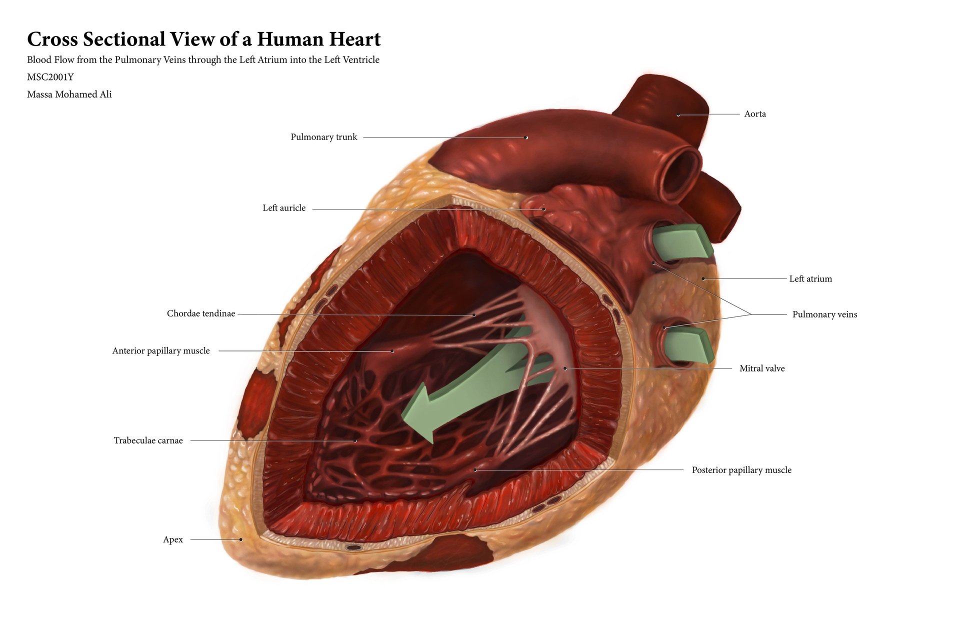
Cross Sectional View of a Human Heart
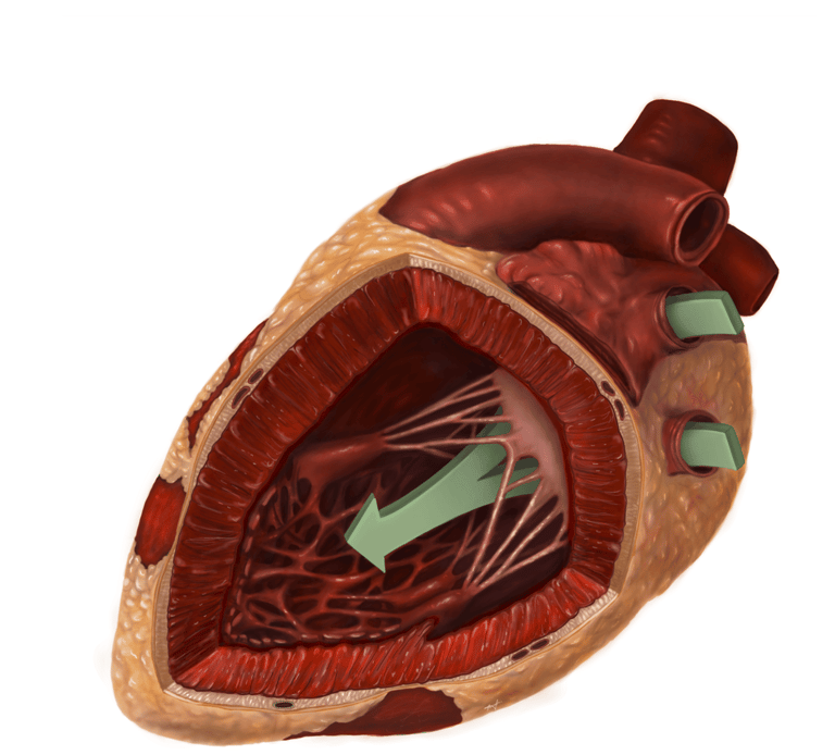
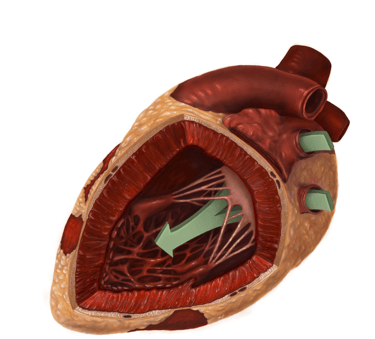
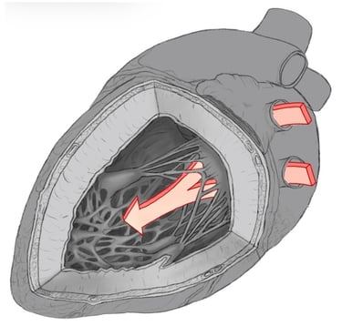
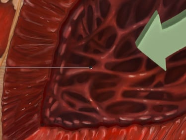
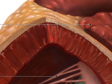
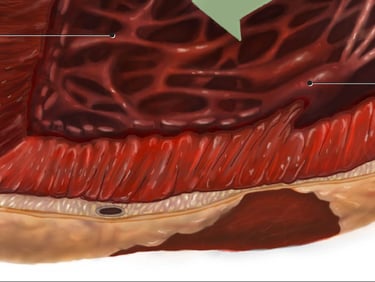
A full colour rendering of a cross-sectional view of a human heart that has not been seen before and could not be seen in an atlas or in Grant’s Museum, created with a high degree of structural accuracy suggesting form, volume, and texture and making the organ look alive. This view shows how blood travels from the pulmonary veins through the left atrium and into the left ventricle.
Softwares used: Adobe Illustrator, Procreate, Cinema4D, ZBrush
Audience: First year anatomy students
Intended Use: Anatomy textbooks
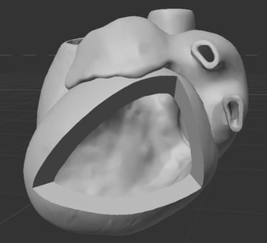
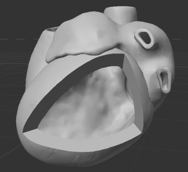
1. Research & Maquette Building
To start this project, I first built a maquette of the human heart in Cinema4D with the intended view and orientation of the cut plane. (Source: Anatomography)
Before developing initial sketches for this piece, I dedicated extensive time researching heart anatomy and blood flow through the left atrium into the left ventricle. My sources included cardiac surgery textbooks and primary anatomical research literature.
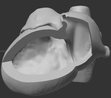
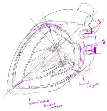
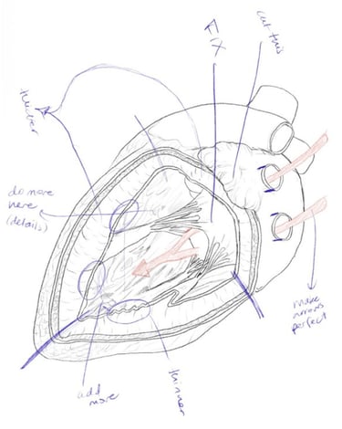
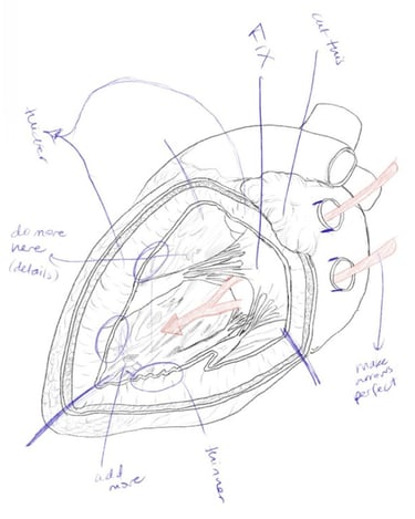
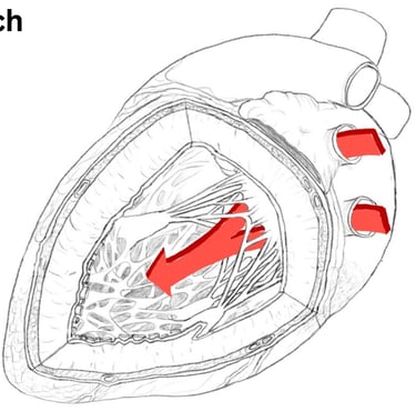
2. Initial Sketches & Revisions
I began with a series of initial sketches to explore the composition, anatomy, and overall structure of the heart. I then revised and refined these concepts through multiple rounds of development before creating the final sketch.
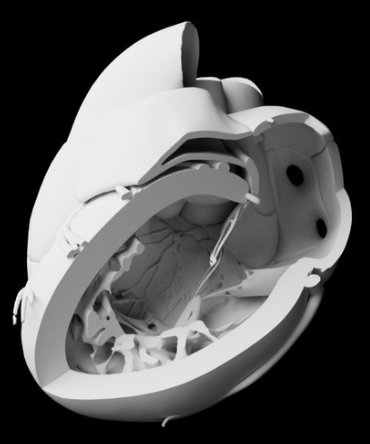
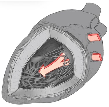
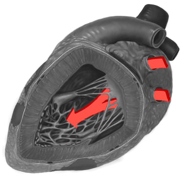
3. Greyscale Rendering
I created a grayscale rendering to establish the full range of tonal values within the piece. These values later served as the foundation for the colourization stage. I paid particular attention to the texture of the trabeculated muscle fibers, rendered the striations along the cut surface of the myocardium, and added highlights to enhance depth and realism.
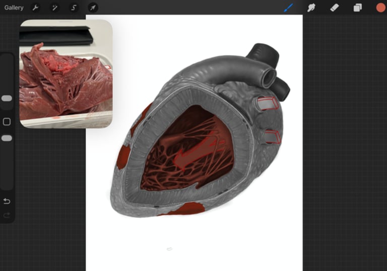
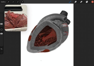
4. Adding Colour
I applied colour gradually in layers, carefully refining the hues and values at each stage to achieve a cohesive and realistic final rendering. Throughout the process, I referenced anatomical textbook images as well as observations from our pig heart dissection to ensure anatomical accuracy and realism.


5. Details & Final Revisions
I finalized the piece by enhancing highlights and textures and adding labels to clearly identify the major structures. I drew in the guiding arrows from the initial sketch and tested different colour combinations to achieve optimal contrast and visual clarity. I also added diffuse and specular highlights to enhance the realism of the tissue.
References
Acland, R. D. (n.d.). Acland’s video atlas of human anatomy [Video]. Lippincott Williams & Wilkins.
Agur, A. M. R., & Dalley, A. F. (2017). Grant’s atlas of anatomy (14th ed.). Wolters Kluwer Health.
Berdajs, D. (n.d.). Operative anatomy of the heart. Springer.
BioDigital. (n.d.). Male adult heart blood flow [3D model]. https://human.biodigital.com/view? id=production/maleAdult/heart_blood_flow&lang=en
Hurst, J. W. (n.d.). Atlas of the heart. McGraw-Hill.
Kenhub. (n.d.). Layers of the heart: Epicardium, myocardium, endocardium. https://www.kenhub.com/en/library/anatomy/layers-of-the-heart
MitralValveRepair.org. (n.d.). Papillary muscles and left ventricle. https://www.mitralvalverepair.org/papillary-muscles-and- left-ventricle
Moore, K. L., Agur, A. M. R., & Dalley, A. F. (2018). Moores Essential Clinical Anatomy (6th ed.). Wolters Kluwer.
National Institutes of Health. (n.d.). 3D Print Exchange: Heart model [3D model]. https://3d.nih.gov/entries/2636
Netter, F. H. (n.d.). Netter atlas of anatomy. Elsevier.
Photographic Reference of the Human Body. (n.d.). Photographic reference of the human body. Publisher.
SciePro. (n.d.). Medical illustrations. https://www.sciepro.com/illustrations?page=37
Sketchfab. (n.d.). 3D heart model [3D model]. https://sketchfab.com/models/a3f0ea2030214a6bbaa97e7357eebd58/embed
StatPearls Publishing. (n.d.). Anatomy, thorax, heart (StatPearls). https://www.ncbi.nlm.nih.gov/books/NBK545203/
The Database Center for Life Science. (n.d.). BodyParts3D/Anatomography: Select parts and make embeddable model of your own [Anatomical 3D database]. Life Science Database Integration Project. https://lifesciencedb.jp/bp3d/?lng=en
University of Minnesota Visible Heart Laboratory. (n.d.). Left ventricle and mitral valve. https://www.vhlab.umn.edu/atlas/left-ventricle/mitral-valve/index.shtml
Vigué-Martínez, A. (n.d.). 248 colour atlas of anatomy. Igaku-Shoin.